Foot X Ray Anatomy - Foot annotated x-ray | Image | Radiopaedia.org / Submitted on march 27, 2012.. The foot and ankle can be subdivided into 4 different parts: It is performed to look for evidence of injury (or pathology) affecting the foot, often after trauma. To download this image, create an account. The ankle joint, tendons of the ankle joint foot anatomy vector illustration. Normal foot and ankle x ray anatomy:
Hover on/off image to show/hide findings. Osteoporosis, a condition in which the bones become weak and brittle, is detectable on an. Another view that is commonly done in addition to these views is lateral view. There are more than a hundred muscles, tendons and ligaments. Xray of the foot is ordered in various ailments of the foot including trauma.
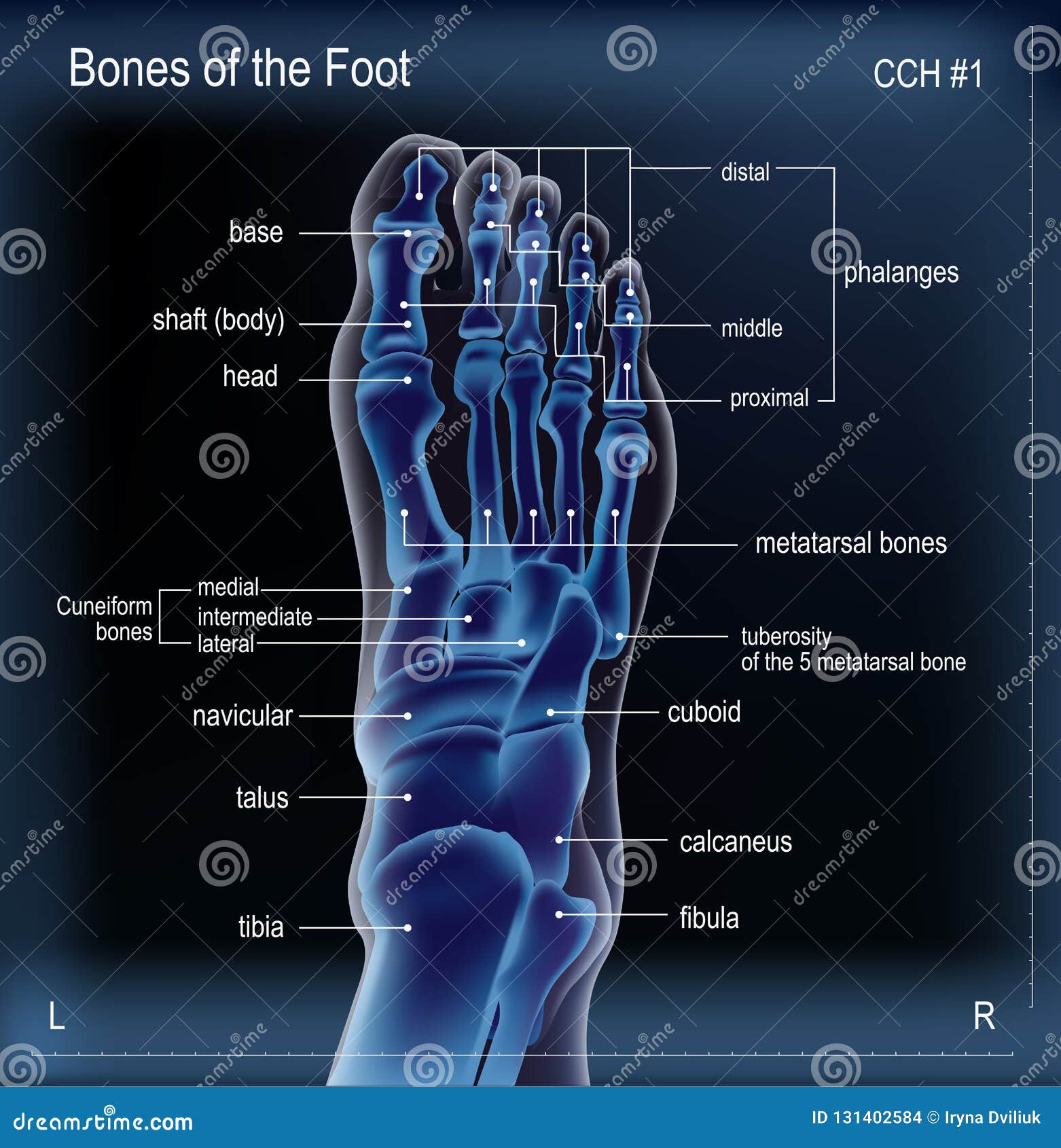
X ray of bones the of foot stock vector. Illustration of ... from thumbs.dreamstime.com Webmd's feet anatomy page provides a detailed image and definition of the parts of the feet and explains their function. The tibia and fibula make up just above the ankle. Metatarsals and phalanges of the toes are numbered 1 to 5. Anteroposterior and oblique views are most common views that are ordered on the foot. Osteoporosis, a condition in which the bones become weak and brittle, is detectable on an. It can help you understand our world more detailed and specific. Presentation outline • relevant anatomy • x ray positioning • interpretation of x rays • lines and angles • relevant pathology. • central ray perpendicular through the ankle joint at a point midway between the.
Anteroposterior and oblique views are most common views that are ordered on the foot.
There is a printable worksheet available for download here so you can take. Normal foot and ankle x ray anatomy: Can also be used for. We think this is the most useful anatomy picture that anatomy is the amazing science. Metatarsals and phalanges of the toes are numbered 1 to 5. It contains information about the normal anatomy and the most common pathology. Osteoporosis, a condition in which the bones become weak and brittle, is detectable on an. Anterior communicating artery anterior cerebral artery rt mri. Flex the ankle and foot enough to place the long axis of the foot in the vertical position. The foot and ankle can be subdivided into 4 different parts: Another view that is commonly done in addition to these views is lateral view. Imaging techniques are able to provide further data on functional anatomy of the foot; Foot anatomy bones lower superior radiograph limb imaios arteries radiology human joints anatomical medical imaging rays labeled extremity atlas feet.
Presentation outline • relevant anatomy • x ray positioning • interpretation of x rays • lines and angles • relevant pathology. In particular, these techniques show the bones structures, ligaments, muscles and tendons, taking part to the arch setting. Anteroposterior and oblique views are most common views that are ordered on the foot. Osteoporosis, a condition in which the bones become weak and brittle, is detectable on an. Metatarsals and phalanges of the toes are numbered 1 to 5.
The 8 best Foot x ray images on Pinterest | Human anatomy ... from i.pinimg.com It can help you understand our world more detailed and specific. The feet are flexible structures of bones, joints, muscles, and soft tissues that let us stand upright and perform activities like walking, running, and jumping. The rear foot, the mid foot, and the forefoot: Foot anatomy bones lower superior radiograph limb imaios arteries radiology human joints anatomical medical imaging rays labeled extremity atlas feet. Can also be used for. We hope you will use this picture in the study and. Superior radiograph of the foot with all anatomical. It is performed to look for evidence of injury (or pathology) affecting the foot, often after trauma.
Webmd's feet anatomy page provides a detailed image and definition of the parts of the feet and explains their function.
Anatomy note anatomy is a great science. The feet are flexible structures of bones, joints, muscles, and soft tissues that let us stand upright and perform activities like walking, running, and jumping. Hover on/off image to show/hide findings. The tibia and fibula make up just above the ankle. The ankle joint, tendons of the ankle joint foot anatomy vector illustration. Superior radiograph of the foot with all anatomical. We hope you will use this picture in the study and. Check you have the right views. Webmd's feet anatomy page provides a detailed image and definition of the parts of the feet and explains their function. Can also be used for. Vintage anatomy print of the human foot, showcasing the veins and arteries. It contains information about the normal anatomy and the most common pathology. Flex the ankle and foot enough to place the long axis of the foot in the vertical position.
There are more than a hundred muscles, tendons and ligaments. The foot and ankle can be subdivided into 4 different parts: Anterior communicating artery anterior cerebral artery rt mri. The rear foot, the mid foot, and the forefoot: Anteroposterior and oblique views are most common views that are ordered on the foot.
8 best Foot x ray images on Pinterest | Human anatomy ... from i.pinimg.com Normal foot and ankle x ray anatomy Flex the ankle and foot enough to place the long axis of the foot in the vertical position. In particular, these techniques show the bones structures, ligaments, muscles and tendons, taking part to the arch setting. The foot and ankle can be subdivided into 4 different parts: The feet are flexible structures of bones, joints, muscles, and soft tissues that let us stand upright and perform activities like walking, running, and jumping. Another view that is commonly done in addition to these views is lateral view. Anatomy note anatomy is a great science. Anteroposterior and oblique views are most common views that are ordered on the foot.
It is performed to look for evidence of injury (or pathology) affecting the foot, often after trauma.
Imaging techniques are able to provide further data on functional anatomy of the foot; Another view that is commonly done in addition to these views is lateral view. To download this image, create an account. There are more than a hundred muscles, tendons and ligaments. Normal foot and ankle x ray anatomy It is performed to look for evidence of injury (or pathology) affecting the foot, often after trauma. Superior radiograph of the foot with all anatomical. There is a printable worksheet available for download here so you can take. In fact every radiologst should be an expert in chest film reading. Metatarsals and phalanges of the toes are numbered 1 to 5. Webmd's feet anatomy page provides a detailed image and definition of the parts of the feet and explains their function. In particular, these techniques show the bones structures, ligaments, muscles and tendons, taking part to the arch setting. Check you have the right views.


Belum ada Komentar untuk "Foot X Ray Anatomy - Foot annotated x-ray | Image | Radiopaedia.org / Submitted on march 27, 2012."
Posting Komentar